14+ areolar tissue diagram
Which structuretissue protects the plants body against the invasion of parasites. 05142021 Table of Contents.

How To Draw Areolar Tissue Diagram Diagram Of Areolar Tissue In Simple Easy Way Areolar Tissue Youtube
C Tissue that transports food in plants.
. Jelly-Like Connective Tissue 9. B Tissue that connects muscle to bone in humans. Draw the diagram for each type of epithelial tissue.
Connective tissue is one of the four main tissue types. Loose Connective Tissue 2. There are two main types of nervous tissue neurons and neuroglia.
Yellow Elastic Tissue 5. INTEXT Q3 PG 74 4. How does a cork act as a protective tissue.
Differentiate the following activities on the basis of voluntary or involuntary. Explore the fibrous tissue of the human body- learn what connective tissues function is the types of fibrous connective tissue and how it supports the body. Brown adipose tissue is for thermal heat regulation.
Write a short note on epithelial tissue. Learn about the structure location and function of dense regular connective tissue in the human body with histological photos and diagrams. Mention the function of the following.
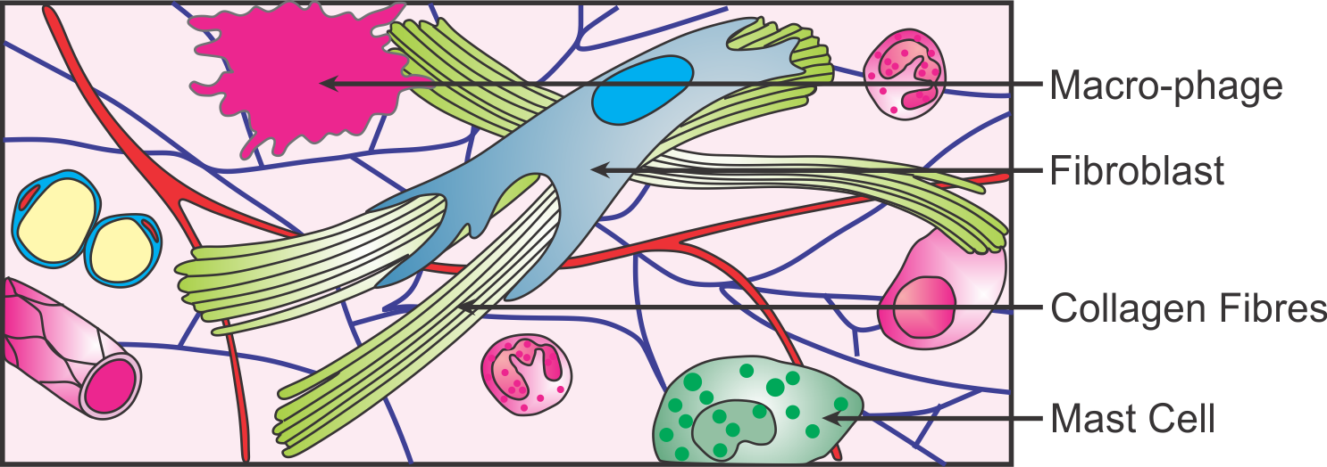
Connective tissue includes tissues such as cartilage bone blood and fat. This diagram shows from left to right. Areolar connective tissue helps in the repair of tissue.
Areolar tissue a connective tissue. Diagram of a neuron along with the labelling is as follows. Enter the email address you signed up with and well email you a reset link.
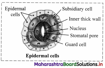
Draw a diagram of leaf epidermal peel showing stomata and label its parts. State the characteristics of cells of epidermis. Single long part of nerve cell.
Physiology Function of the Integumentary System. Epithelial tissues are of following types. It occurs inside organs around blood vessels muscles and nerves below skin subcutaneous tissue and joining various structures like muscles with skin.
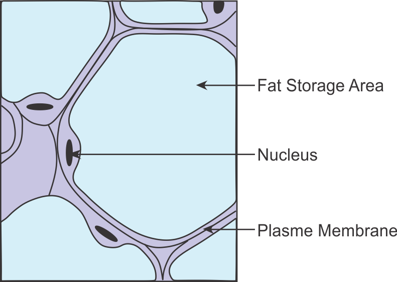
Nervous tissue connective tissue muscle tissue and epithelial tissue. White Fibrous Tissue 4. Loose areolar connective tissue and adipose tissue which functions as a mode of fat storage and provides insulation and cushioning for the integument.
Which muscle has spindle-shaped cells. Learn how areolar connective tissue differs from other tissue types. Types of Nervous Tissue.
Smooth muscle cells have spindle shaped cells. Areolar connective tissue is the most abundant of the connective tissues in the body. Movement of passage of food in the intestine is caused by the contraction of a cardiac muscles b unstriated muscles.
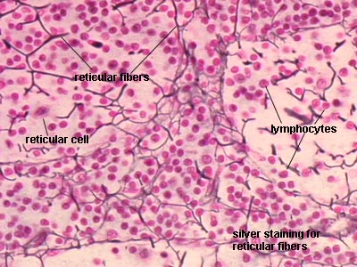
F Tissue present in the brain. Blood and Haemopoietic Tissue 7. A Ureters in frog b Malpighian tubules c Body wall in earthworm.
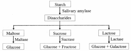
What are the functions of areolar tissue. These two types of cells fulfill different roles in the nervous system and can be found in different. The gastrointestinal tract GI tract digestive tract alimentary canal is the tract or passageway of the digestive system that leads from the mouth to the anusThe GI tract contains all the major organs of the digestive system in humans and other animals including the esophagus stomach and intestinesFood taken in through the mouth is digested to extract nutrients and absorb.
E Differences between simple gland and compound gland are as follows. As you may have learned there are four types of tissues that compose all of the structures of our bodies. Though nearly all human skin is covered with hair follicles it can appear.
Osseous Tissue or Bone 10. The following points highlight the ten main varieties of connective tissues of human body. Areolar tissue is a connecting tissue found between skin and muscles around our.
It is sturdier than adipose tissue but still flexible. Describe the functions of epithelium tissue. Muscle tissue nervous tissue connective tissue - each one serves a purpose within the body through the transmission of messages movement and the protection of organs.
This diagram shows areolar connective tissue serving as a medium between epithelial. Draw a labelled diagram of unstriated muscle tissue and mention its occurrence features and. Areolar and adipose tissue areolar connective tissue areolar connective tissue diagram areolar connective tissue function areolar connective tissue joins areolar connective tissue labeled areolar connective tissue location areolar connective tissue would be.
A Simple squamous epithelium. A Draw diagram of neuron and label the following points on it. Dense Connective Tissues 3.
Figure 514 image description. A Areolar Tissue Loose connective tissue. Adipose tissue helps in storage of fats and acts as heat insulator.
The others are epithelial tissue muscle tissue and nervous tissue. A Tissue that forms the inner lining of our mouth. E Connective tissue with a fluid matrix.
On the diagram above find the two layers of the skin. What is a goblet cell. This tissue is most widely distributed connective tissue in the animal body.
The tissue that helps in the secretion and absorption and is found in the inner lining of the alimentary canal is a ciliated epithelium b cuboidal epithelium. D Tissue that stores fat in our body. Location and Diagram.
The human skin is the outer covering of the body and is the largest organ of the integumentary systemThe skin has up to seven layers of ectodermal tissue guarding muscles bones ligaments and internal organsHuman skin is similar to most of the other mammals skin and it is very similar to pig skin. The epithelial tissue made up of a single layer of epithelial cells of different heights is known as the pseudostratified columnar epitheliumThe position of. Loose connective tissue white adipose tissue and fibrous connective tissue.
Pseudo-Stratified Columnar Epithelium. The following points highlight the three main types of connective tissues. D Differences between adipose tissue and blood tissue are as follows.
10192021 Table of Contents. Areolar tissue is the most widely distributed solid connective tissue in the body. Draw a neat diagram of digestive system of frog.
Draw a labelled diagram of neuron. 1 Tissue that forms the inner lining of our mouth. Draw a well labeled diagram of neuron.
Practice Labeling the Layers of the Skin.

Western Medical Research Conference Formerly Western Regional Meeting January 25 27 2018 Carmel California Journal Of Investigative Medicine

Essential Role Of Elovl4 Protein In Very Long Chain Fatty Acid Synthesis And Retinal Function Journal Of Biological Chemistry
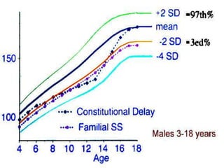
Approach To Short Stature

A Graft Of Perifascial Areolar Tissue Pat A A Layer Of Loose Download Scientific Diagram
Class 11 Page 22 Balbharati Solutions

Areolar Tissue Diagram Google Search Tissue Types Tissue Collagen Fibers

Areolar Connective Tissue Function And Components Jotscroll

Draw A Labeled Diagram Of Areolar Tissue Brainly In

Areolar Connective Tissue Function Location What Is Areolar Connective Tissue Video Lesson Transcript Study Com

The Structure And Function Of The Cervix During Pregnancy Sciencedirect
Proper Connective Tissue Areolar Adipose Reticular White Fibrous And Yellow Elastic Tissue Online Biology Notes
Draw A Labelled Diagram Of Areolar Connective Tissue Sarthaks Econnect Largest Online Education Community

Essential Role Of Elovl4 Protein In Very Long Chain Fatty Acid Synthesis And Retinal Function Journal Of Biological Chemistry

How To Draw Areolar Tissue Diagram Diagram Of Areolar Tissue In Simple Easy Way Areolar Tissue Youtube
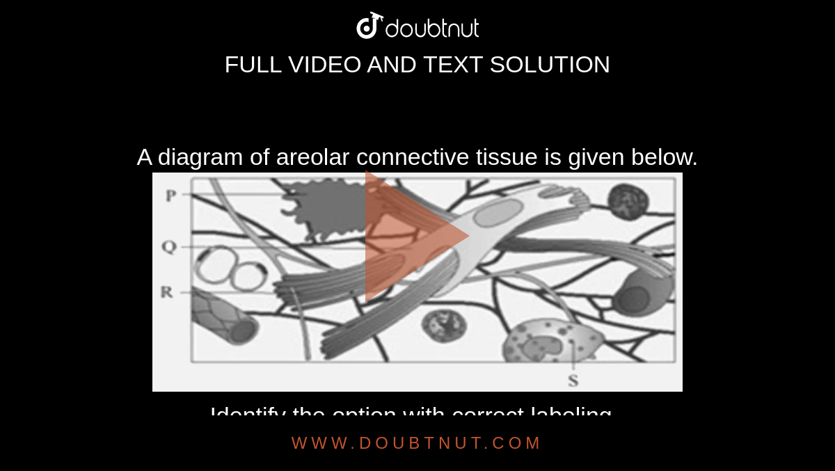
A Diagram Of Areolar Connective Tissue Is Given Below Identify The Option With Correct Labeling
Cbse Class 9 Answered
Bhagya Page 62 Balbharati Solutions